Why “Normal” Heart Tests Do Not Always Mean Your Coronary Arteries Are Clear
“My ECG is normal.”
“My echo is normal.”
“So there cannot be any blockage in my heart… right?”
This is one of the most common—and understandable—questions patients ask a cardiologist.
A normal ECG and a normal echocardiogram are certainly reassuring findings.
But they do not automatically rule out coronary artery disease.
This becomes especially important when someone continues to experience symptoms such as:
- Chest pressure while walking
- Tightness in the chest
- Breathlessness on exertion
- Reduced walking capacity
- Symptoms that disappear after resting
In such situations, the symptoms may be telling the doctor something that a resting ECG or echo cannot show.
Dr. S. Nagendra Boopathy explains this using a real clinical situation involving one of his patients.
A 70-Year-Old Patient Who Looked “Normal” on Tests
The patient was around 70 years old.
He had several cardiovascular risk factors:
- Diabetes
- High blood pressure
- High cholesterol
He had been taking treatment for approximately 10 years, and importantly, his conditions appeared reasonably well controlled.
He had previously been an active person and could walk comfortably.
But over the previous six months, something had changed.
Whenever he walked, he started experiencing:
- Mild breathlessness
- A feeling of heaviness or tightness in his chest
- A pressure-like sensation
When he stopped walking and rested, the symptoms improved.
Initially, he was still able to walk reasonably well.
But during the following months, his exercise capacity progressively declined.
Then, during the final couple of weeks before seeing the cardiologist, the symptoms became much more significant.
He could barely walk a short distance before the chest pressure appeared.
That change was important.
“But Doctor, My Sugar, BP and Cholesterol Are Controlled”
During the consultation, the patient’s blood pressure was controlled.
His cholesterol treatment appeared effective.
His diabetes was being managed.
But Dr. Boopathy was concerned about the patient’s symptoms.
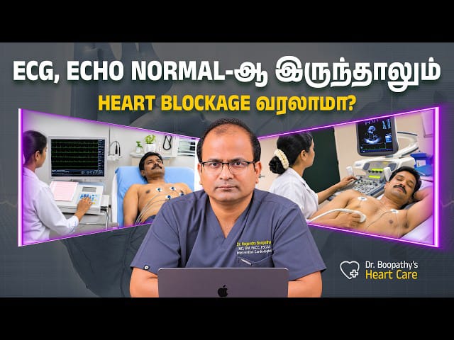
So an ECG and an echocardiogram were performed.
And both were largely reassuring.
ECG: Normal
Heart pumping function: Normal
Major valve function: Normal
The echo showed only relatively mild changes that could be related to longstanding high blood pressure, including thickening of the heart muscle and some degree of impaired relaxation.
There was no major pumping abnormality.
The patient and his wife naturally felt relieved.
But the cardiologist still said:
“Your symptoms make me suspicious that there may be a problem with blood flow through the coronary arteries.”
That surprised them.
Their question was immediate:
“If the ECG and echo are normal, how can there still be a heart problem?”
To understand the answer, we first need to understand what each test actually measures.
What Exactly Is an ECG?
ECG stands for:
Electrocardiogram
Your heart is not simply a mechanical pump.
It also has its own electrical system.
Every heartbeat begins with an electrical impulse.
The signal normally starts in the heart’s natural pacemaker—the sinoatrial or SA node.
It then travels through the electrical pathways of the heart, including the:
- AV node
- His-Purkinje system
- Right bundle branch
- Left bundle branch
These signals coordinate the contraction of the heart muscle.
An ECG records this electrical activity.
What Can an ECG Tell Us?
An ECG can provide important information about:
Heart rhythm
Is the heartbeat regular or irregular?
Heart rate
Is it too slow or too fast?
Electrical conduction
Are electrical signals travelling normally through the heart?
Previous heart muscle damage
Certain patterns may suggest a previous heart attack.
Acute heart injury
If a person is having certain types of heart attack while the ECG is being recorded, characteristic changes may appear.
Enlargement or thickening
Certain ECG patterns may suggest enlargement or thickening of heart chambers.
But there is one important limitation.
A Resting ECG Is Only a Snapshot
An ordinary ECG usually records the heart for only a short period of time.
And most commonly, it is recorded when the person is:
- Lying down
- Resting
- Not exercising
- Not experiencing significant physical stress
So a normal resting ECG essentially tells us:
“At this particular moment, the electrical activity of the heart does not show the abnormalities this test is designed to detect.”
That is useful.
But it does not mean:
“There is definitely no cholesterol plaque or narrowing in the coronary arteries.”
These are different questions.
“Heart Block” and “Heart Artery Blockage” Are Not the Same Thing
This causes enormous confusion among patients.
Someone may say:
“The doctor told me I don’t have a heart block.”
But what kind of “block” are we talking about?
There are two completely different conditions.
Electrical Heart Block
This means there is a problem with how electrical signals travel through the heart.
An ECG is particularly useful for detecting this.
Coronary Artery Blockage
This means narrowing or obstruction in the blood vessels supplying oxygen-rich blood to the heart muscle.
This usually develops because of atherosclerotic plaque.
An ECG does not directly show the coronary arteries.
Therefore:
A normal ECG does not automatically mean your coronary arteries are normal.
Then What Is an Echocardiogram?
An echocardiogram—or “echo”—uses ultrasound waves to create moving images of the heart.
Many people are already familiar with ultrasound because it is commonly used during pregnancy.
The same basic imaging principle can be used to look at the heart.
An echo allows the cardiologist to evaluate the heart while it is beating.
What Does an Echo Show?
An echocardiogram can help doctors assess:
Heart pumping function
How strongly is the heart contracting?
Heart muscle movement
Are different areas of the heart moving normally?
Chamber size
Are any heart chambers enlarged?
Heart muscle thickness
Is the muscle thickened, as can occur with longstanding hypertension?
Heart valves
Are the valves opening and closing normally?
Is there narrowing or leakage?
Congenital abnormalities
Are there structural abnormalities such as certain holes or abnormal connections?
Fluid around the heart
Is there abnormal fluid in the sac surrounding the heart?
It is an extremely useful test.
But again, a routine resting echo has limitations.
A Normal Echo Does Not Necessarily Mean Your Arteries Are Clear
An echo may show:
- Normal pumping
- Normal valve function
- Normal chamber sizes
- No obvious major heart muscle damage
And yet a patient can still have coronary artery narrowing.
Why?
Because the heart may receive enough blood while the body is resting.
The problem may only become apparent when the heart has to work harder.
This is where the difference between rest and stress becomes important.
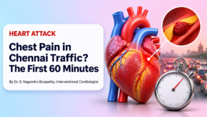
Think About Chennai Traffic
Dr. Boopathy uses a simple example.
Imagine travelling from Porur to Kodambakkam.
If you travel during a relatively quiet time of the day, the journey may take around 20–30 minutes.
The road appears perfectly manageable.
But suppose you travel during evening rush hour.
Suddenly:
- Traffic increases
- Roads become congested
- The same route takes much longer
The road did not suddenly become a different road.
The problem became obvious only when the demand increased.
The heart can behave in a similar way.
Your Heart at Rest vs Your Heart During Exercise
When you are lying quietly on a bed:
- Heart rate is lower
- Blood pressure demand may be lower
- Heart muscle requires less oxygen
Even a coronary artery that is partially narrowed may still deliver enough blood for this resting state.
Therefore:
Resting ECG → may look normal
Resting echo → may look normal
But now imagine you start walking briskly.
Your muscles need more oxygen.
Your heart beats faster.
The heart muscle itself needs more blood.
If there is a significant narrowing in a coronary artery, blood flow may not increase enough to meet this demand.
That is when symptoms may appear.
What Symptoms Suggest Reduced Blood Flow to the Heart?
Symptoms may include:
- Chest pressure
- Chest tightness
- Heaviness
- Squeezing discomfort
- Breathlessness
- Unusual fatigue
Some people describe the feeling as:
“My chest feels compressed.”
Others may not describe it as pain at all.
They may simply say:
“When I walk, I feel uncomfortable in my chest. When I stop, it disappears.”
That pattern matters.
Why Symptoms That Occur During Walking Are Important
In the patient discussed by Dr. Boopathy, the symptoms were strongly related to exertion.
He would walk.
Symptoms would appear.
He would stop.
Symptoms would settle.
Then the amount of activity required to trigger symptoms became progressively smaller.
Initially he could walk much farther.
Later, even a short distance caused chest pressure.
That worsening pattern deserves careful evaluation regardless of whether the resting ECG and echo look reassuring.
What Happens Next If ECG and Echo Are Normal?
The answer is not:
“Everyone needs more tests.”
This is one of Dr. Boopathy’s most important messages.
Not every person with a normal ECG needs:
- A treadmill test
- CT coronary angiography
- Nuclear imaging
- Angiography
Testing should be based on the patient’s:
- Symptoms
- Age
- Risk factors
- Examination
- Medical history
- Existing test results
- Overall likelihood of coronary artery disease
The right patient should undergo the right test at the right time.
What Is a Stress Test?
A stress test evaluates how the heart behaves when its workload increases.
Instead of examining the heart only while the patient is quietly resting, doctors deliberately increase the heart’s workload.
This can sometimes uncover abnormalities that were not visible at rest.
1. Treadmill Exercise Test
During a treadmill test, the patient walks on a treadmill while healthcare professionals monitor:
- ECG
- Heart rate
- Blood pressure
- Symptoms
The speed and incline gradually increase according to a standard protocol.
The aim is to see whether exertion causes:
- Typical symptoms
- Significant ECG changes
- Abnormal blood-pressure response
- Rhythm abnormalities
A treadmill test can be useful in selected patients who are able to exercise and have an interpretable resting ECG.
It is not the best test for every patient.
2. Stress Echocardiography
Another option is stress echocardiography.
The heart is evaluated using ultrasound before and during or immediately after stress.
Stress may come from exercise or, in selected cases, medication that makes the heart work harder.
If part of the heart muscle does not receive adequate blood during stress, its movement can sometimes become abnormal.
That can provide evidence of myocardial ischemia.
3. Medication-Induced Stress Testing
Not every patient can exercise adequately on a treadmill.
For example, someone may have:
- Severe arthritis
- Walking difficulty
- Neurological problems
- Poor exercise capacity
In selected situations, medication can be used to increase the heart’s workload or alter coronary blood flow.
One example discussed by Dr. Boopathy is dobutamine, which increases heart rate and contractility and can be used during stress echocardiography.
4. Nuclear Stress Testing
Nuclear perfusion tests can evaluate blood flow to the heart muscle.
These tests may use tracers and imaging methods such as myocardial perfusion imaging.
The goal is to identify whether a particular region of heart muscle receives less blood during stress than at rest.
5. CT Coronary Angiography
CT coronary angiography—often called CCTA—takes a different approach.
Instead of primarily looking for the effect of reduced blood flow, CT imaging can provide detailed anatomical pictures of the coronary arteries.
It may show:
- Coronary plaque
- Location of narrowing
- Extent of disease
- Whether significant obstructive coronary disease may be present
For appropriately selected patients, CT coronary angiography can be extremely useful.
Does Everyone With Chest Pain Need a Stress Test?
No.
This is important.
Chest discomfort has many possible causes.
Some people have a very low likelihood of coronary artery disease.
Others may need different investigations.
Modern cardiovascular assessment increasingly uses structured clinical risk evaluation to decide who is most likely to benefit from additional testing.
The decision should be individualised.
Why Are Diabetes, BP and Cholesterol Important Even When Controlled?
The patient in Dr. Boopathy’s example had:
- Diabetes
- Hypertension
- Cholesterol problems
His numbers were currently well controlled.
That is excellent.
Treatment reduces cardiovascular risk.
But good control today does not erase all the cumulative effects of having had cardiovascular risk factors over many years.
Atherosclerosis develops gradually.
This is why doctors consider both:
What your numbers are today
and
Your long-term medical history.
Can Someone With Good Cholesterol Still Have Coronary Disease?
Yes.
Cholesterol is extremely important, but cardiovascular risk is influenced by several factors.
These include:
- Age
- Diabetes
- Blood pressure
- Smoking
- Cholesterol exposure over time
- Family history
- Kidney disease
- Physical inactivity
- Obesity
- Other metabolic factors
A single “normal” report cannot tell the entire story.
What Is Mild Heart Muscle Thickening on Echo?
Longstanding high blood pressure can make the heart pump against increased resistance.
Over time, the main pumping chamber—the left ventricle—may become thicker.
This is often referred to as left ventricular hypertrophy.
If thickening is relatively uniform, it may be described as concentric hypertrophy.
The heart can still have normal squeezing strength while becoming somewhat stiffer.
What Is Diastolic Dysfunction?
The heart has two broad phases:
Systole
The heart contracts and pumps blood out.
Diastole
The heart relaxes and fills with blood.
When the heart muscle becomes thicker or stiffer, relaxation can become less efficient.
This may be described as diastolic dysfunction.
Mild diastolic dysfunction can occur with:
- Ageing
- Hypertension
- Diabetes
- Other cardiovascular conditions
It does not automatically explain every episode of exertional chest pressure.
The full clinical picture still matters.
Can a Heart Attack Occur With a Previously Normal ECG?
Yes.
An ECG represents what is happening during the period when it is recorded.
A normal ECG weeks or months ago does not guarantee that a new cardiac event cannot develop later.
Even during an acute evaluation, a single normal ECG does not by itself exclude every form of acute coronary syndrome.
Symptoms and clinical risk remain important.
Should I Ignore Chest Pain If My Previous Tests Were Normal?
No.
This is perhaps the most important practical message from this discussion.
If you develop:
- New chest pressure
- Increasing chest tightness
- Breathlessness on exertion
- A significant reduction in walking capacity
- Symptoms that are becoming more frequent
you should seek medical assessment.
Do not reassure yourself simply by saying:
“But my ECG was normal last year.”
The situation today may be different.
When Is Chest Pain an Emergency?
Do not wait for an outpatient appointment if you develop severe or persistent symptoms such as:
- Chest pressure at rest
- Chest discomfort lasting several minutes or repeatedly returning
- Severe breathlessness
- Cold sweating
- Fainting
- Sudden weakness
- Chest discomfort with nausea or extreme uneasiness
- Pain or pressure spreading to the arm, shoulder, jaw or back
Seek urgent emergency medical attention.
The Most Important Lesson: Tests Should Follow the Patient, Not the Other Way Around
Medicine is not simply about ordering every available investigation.
Imagine two people.
Patient A
Young, no cardiovascular risk factors, no symptoms, normal examination.
Patient B
Older, diabetic, hypertensive, with progressively worsening exertional chest pressure.
Both may have a normal resting ECG.
But their clinical situations are completely different.
Therefore, the next step should not be determined by the ECG result alone.
A good clinical evaluation considers:
- The patient’s story
- Symptoms
- Risk factors
- Examination
- Existing investigations
and then chooses the investigation most likely to answer the clinical question.
As Dr. Boopathy summarises:
The right test, for the right patient, at the right time.
Frequently Asked Questions
1. Can I have a heart blockage even if my ECG is normal?
Yes.
A resting ECG mainly records the heart’s electrical activity during that particular period. It does not directly show the coronary arteries.
2. Does a normal ECG mean my heart is completely healthy?
A normal ECG is reassuring, but it cannot rule out every heart condition.
Its interpretation should be combined with your symptoms, examination and risk factors.
3. Does an ECG show cholesterol blockage?
Not directly.
An ECG can sometimes show changes caused by reduced blood flow or heart muscle injury, but it does not provide an image of cholesterol plaque inside an artery.
4. Is “heart block” the same as coronary artery blockage?
No.
Electrical heart block refers to abnormal electrical conduction.
Coronary artery blockage refers to narrowing in the vessels carrying blood to the heart muscle.
They are different problems.
5. Can an echo detect coronary artery blockage?
A standard resting echo generally does not directly visualise coronary artery narrowing.
It assesses heart structure, pumping, valves and heart muscle movement.
6. If my echo pumping function is normal, can I still have coronary artery disease?
Yes.
A person can have normal pumping function and still have significant coronary artery disease.
7. What does an echo actually look for?
It can assess heart pumping, chamber size, muscle thickness, valve function, structural abnormalities and fluid around the heart.
8. Why can heart tests be normal at rest but abnormal during exercise?
The heart requires much less oxygen while resting.
During exercise, demand increases.
A narrowed coronary artery may supply enough blood at rest but fail to meet the increased demand during exertion.
9. What is angina?
Angina is discomfort caused by inadequate blood supply to heart muscle.
It may feel like:
- Pressure
- Tightness
- Heaviness
- Squeezing
- Breathlessness
It does not always feel like sharp pain.
10. Why does chest pressure improve when I stop walking?
During rest, the heart’s oxygen requirement decreases.
If symptoms are caused by reduced coronary blood flow during exercise, they may improve when the workload falls.
That pattern warrants medical assessment.
11. What is a treadmill test?
It is a monitored exercise test where ECG, heart rate, blood pressure and symptoms are evaluated while the workload gradually increases.
12. Does everyone with chest pain need a treadmill test?
No.
The appropriate investigation depends on age, symptoms, risk factors, exercise ability and other clinical factors.
13. Can older patients undergo treadmill testing?
Some can, provided they can exercise safely and adequately.
Others may benefit from different forms of stress testing or coronary imaging.
14. What if I cannot walk on a treadmill?
Depending on the clinical situation, doctors may use stress imaging with medication or another diagnostic strategy.
15. What is stress echocardiography?
It compares heart function during rest and increased workload to see whether stress produces abnormalities suggestive of reduced blood flow.
16. What is dobutamine stress echo?
Dobutamine is a medication that increases the heart’s workload.
It can be used with echocardiography in selected patients who cannot exercise adequately.
17. What is a nuclear stress test?
It uses a small amount of imaging tracer to evaluate blood flow to heart muscle at rest and during stress.
18. What is CT coronary angiography?
It is a CT-based imaging test that can produce detailed pictures of the coronary arteries and identify plaque or narrowing.
19. Which is better—treadmill or CT angiography?
Neither test is universally “better.”
The correct choice depends on the individual patient and the clinical question being asked.
20. Does everyone with a normal ECG and echo need CT angiography?
No.
Routine additional testing is not necessary for everyone.
Testing should be guided by symptoms and clinical likelihood of coronary artery disease.
21. My cholesterol is controlled. Can I still have a blockage?
Yes.
Good cholesterol control reduces risk, but existing plaque may have developed over many years, and cardiovascular risk involves more than cholesterol alone.
22. My diabetes is controlled. Am I still at risk?
Risk is lower when diabetes is well controlled, but having a history of diabetes remains relevant when doctors assess cardiovascular risk.
23. Can high blood pressure thicken the heart muscle?
Yes.
Longstanding hypertension can cause thickening of the left ventricular muscle.
24. What does diastolic dysfunction mean?
It means the heart muscle does not relax and fill as efficiently as normal.
It is commonly associated with ageing, hypertension and other cardiovascular conditions.
25. What if my ECG last year was normal but I have symptoms now?
You should be evaluated based on your current symptoms.
An old normal test should not be used to dismiss new or worsening symptoms.
26. Can breathlessness be a heart symptom without chest pain?
Yes.
Breathlessness, unusual fatigue or reduced exercise tolerance can sometimes represent a cardiac symptom, particularly in people with cardiovascular risk factors.
27. When should I worry about reduced walking capacity?
If you previously walked comfortably but now have progressively increasing breathlessness, chest pressure or fatigue with smaller amounts of activity, discuss it with a doctor.
28. Does a stress-test abnormality automatically mean I need a stent?
No.
The next step depends on the type and severity of abnormality, symptoms, coronary anatomy and overall clinical condition.
Some patients are treated with medication.
Others may require further investigation or revascularisation.
29. Can a normal stress test guarantee that I will never have heart disease?
No test offers a lifetime guarantee.
Cardiovascular risk changes over time.
Risk-factor control and medical follow-up remain important.
30. What is the most important message from this discussion?
Do not interpret a test result without considering the patient.
A normal resting ECG and echo are reassuring—but persistent or worsening exertional symptoms still deserve proper assessment.
Final Takeaway
Many people believe:
“Normal ECG + normal echo = no heart blockage.”
Unfortunately, it is not that simple.
An ECG primarily tells us about the electrical activity of the heart.
An echocardiogram tells us about heart structure, pumping function and valves.
Both tests are extremely valuable.
But neither is designed to directly rule out every case of coronary artery disease.
Sometimes the heart looks completely normal while resting.
The problem becomes visible only when the heart is asked to work harder.
That is why a patient who develops:
- Pressure while walking
- Breathlessness with exertion
- Progressive reduction in walking capacity
- Symptoms that improve after resting
may need further investigation even when resting tests are reassuring.
The answer is not to test everybody.
Nor is the answer to ignore symptoms because one test is normal.
The correct approach is:
Listen to the symptoms.
Assess the risk.
Choose the right test.
At the right time.
Because in cardiology, the most important test is not always the first test.
It is the test that correctly answers the question raised by the patient’s symptoms.
Consult Dr. S. Nagendra Boopathy
Dr. S. Nagendra Boopathy
MBBS (MMC), MD (PGIMER), DM Cardiology (AIIMS), FACC, FSCAI
Professor of Cardiology & Senior Consultant Cardiologist / Interventional Cardiologist
Dr. Boopathy Heart Care
6th Cross Street
Director’s Colony
Kodambakkam
Chennai – 600024
Appointments:
+91 87544 98680
Sri Ramachandra Medical Centre
No. 1, Ramachandra Nagar
Porur
Chennai – 600116
Appointments: 044-4592 8692
Help Desk: 044-4592 8844 / 044-4592 8855
Hospital Enquiries: 044-4001 2323
Emergency: 044-400 12345
Medical Disclaimer: This article is intended for general educational purposes and does not replace individual medical assessment. A normal resting ECG or echocardiogram cannot independently exclude all forms of coronary artery disease. New, persistent or worsening chest pressure, severe breathlessness, fainting or other symptoms suggestive of a heart attack require prompt medical attention.